Zygomatic Fracture Reconstruction: Customized Approach for Post-Traumatic Deformity
- Dr. Park

- Jan 6, 2025
- 3 min read
Updated: Jan 7, 2025
This is Dr. Jong Chul Park, a board-certified Oral and Maxillofacial Surgeon.
I'd like to share a case of facial bone reconstruction for a patient who suffered a depressed fracture of the left zygoma (cheekbone) and infraorbital rim (bone beneath the eye) due to a motorcycle accident 10 years ago while not wearing a helmet.
This case highlights the potential for depressed deformities following zygomatic fractures and the customized reconstructive approach used to address them.
Depressed Zygoma: Simple Implant Placement May Not Be Sufficient

When a zygomatic fracture occurs, the affected area may appear sunken. This is often due to incomplete reduction (the surgical procedure to reposition fractured bones) during the initial treatment, leading to the bone healing in a misaligned position.
If there are no functional issues, augmenting the deficient volume with an implant can be a viable option with a favorable risk-benefit profile.
However, in cases of trauma to the malar eminence (also known as the 45-degree angle of the cheekbone), while the malar eminence may appear depressed, the lateral zygoma (side of the cheekbone) often becomes wider due to the impact.
Depressed Malar Eminence and Widened Lateral Zygoma: A Complex Problem

In this patient's case, the impact caused depression of the malar eminence, while conversely, the lateral zygoma developed an outward convexity, resulting in a 6mm difference in width between the two sides.
In such situations, simply placing an implant in the depressed malar eminence area could exacerbate the asymmetry by making the left lateral zygoma appear even larger overall.
Precise Diagnosis and Personalized Surgical Planning

This patient required a complex surgical plan that involved restoring the malar eminence laterally while simultaneously reducing the lateral zygoma medially. In other words, a meticulous plan was devised to reposition the entire zygoma and reconstruct the infraorbital depression with a custom implant.
Fabrication of Patient-Specific Bone Repositioning Guide, Customized Plate, and Implant
Fabrication of a guide for patient bone repositioning and customized metal plate: A surgical guide and customized plate were precisely fabricated to reposition the patient's bone to the ideal location.

Fabrication of a guide for patient bone repositioning and customized metal plate Design of patient-specific implant: Using 3D modeling technology, a custom implant was designed to optimally fit the patient's facial structure. In this case, a custom silicone implant was fabricated to effectively restore the depressed area.

Patient-specific zygomatic implant design
Surgical Outcome: Restored Facial Symmetry and Natural Aesthetics


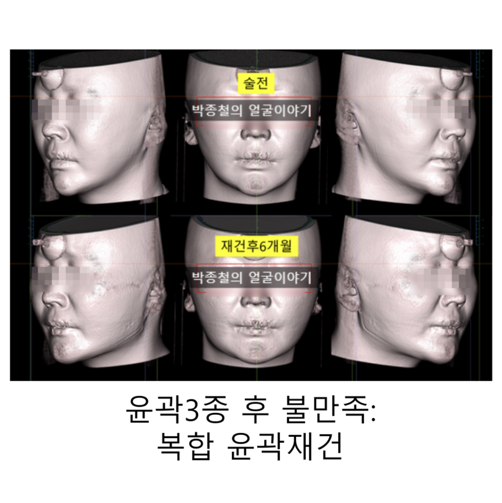
Postoperative CT scans confirmed the successful lateral restoration of the left malar eminence and medial reduction of the lateral zygoma, resulting in restored symmetry. Although the silicone implant is not clearly visible on CT images, pre- and postoperative photographs demonstrate a natural improvement in the depressed area.

Conclusion: Importance of Consulting with a Skilled Specialist for Zygomatic Reconstruction
This case exemplifies the depressed deformities that can occur after zygomatic fractures and the corresponding customized reconstructive approach. Zygomatic reconstruction requires more than just filling a depressed area; it necessitates a precise surgical plan that considers the overall balance and harmony of the face, along with the skills of an experienced specialist. If you are concerned about zygomatic deformities, I encourage you to consult with a board-certified Oral and Maxillofacial Surgeon to obtain an accurate diagnosis and develop a personalized treatment plan.
Viewing the video can help you easily see the changes. Please take a look.


Comments