Zygoma Reduction: A CT Analysis of Cheek Dimpling & Your Eye Contour
- Dr. Park

- Aug 18, 2025
- 3 min read
Hello, I'm Dr. Park Jong-cheol, an Oral and Maxillofacial Surgeon.
When considering zygoma reduction, patients are naturally focused on the final aesthetic result. While clinical before-and-after photos are helpful, they don't tell the whole story. The most accurate way to understand the outcome is to analyze how the foundational changes to the bone directly influence the overlying soft tissue.
Today, we will use a quantitative CT analysis to explore two of the most frequently discussed results of cheekbone surgery: the appearance of cheek dimpling and the subtle but significant changes to your eye contour.
The Foundation: What CT Data Reveals About Surgical Changes
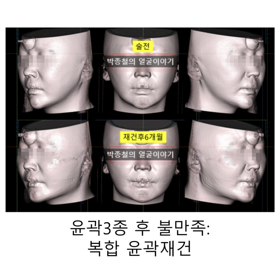
The patient in this case underwent zygoma reduction to soften a prominent 45-degree and lateral zygoma. A CT scan was performed six months after surgery to objectively measure the results.

Our analysis showed a direct correlation between bone movement and soft tissue reduction. For example, when the 45-degree zygoma was moved inward by 4.61mm, the overlying soft tissue contour changed by 3.71mm, meaning about 80% of the bone reduction was reflected in the final facial shape.


This data provides the objective basis for understanding the more nuanced changes discussed below.
How Zygoma Reduction Affects Your Eye Contour
To be clear, the surgery does not alter the eye itself. Instead, it refines your eye contour by changing the structure of the surrounding area.
Our CT analysis revealed soft tissue reduction in the area superior to the bone cut—a region not directly operated on. We measured a soft tissue change of up to 2.28mm in the area near the outer eye.

This happens because reducing the volume of the 45-degree zygoma allows the surrounding soft tissue to settle into a new position. This subtle volumetric change around the socket can make the eyes appear more open or change the overall aesthetic balance of the midface. This is a sophisticated, secondary effect of the surgery that contributes significantly to the final result.

An Analysis of Cheek Dimpling
A primary concern for many patients is the development or accentuation of cheek dimpling after surgery. Our CT analysis provides a clear explanation for this phenomenon.

The key is an anatomical structure called the retaining ligament, a strong fibrous band that connects the cheekbone directly to the skin. When zygoma reduction is performed, the bone is moved inward. Because the skin is tethered to the bone by this ligament, it gets pulled inward as well, which can make a natural dimple more pronounced.

As the CT scan of this patient confirms, the dimple was a pre-existing feature. The surgery did not create it; rather, the reduction of the prominent bone made the effect of this ligament more visible. Understanding this allows us to anticipate this outcome and discuss solutions—such as fat grafting or a mini-lift—for patients who wish to soften the indentation.

Conclusion: An Informed Approach to Surgery
A successful zygoma reduction is one where the patient fully understands the potential outcomes. As this CT analysis demonstrates:
Cheek Dimpling is a result of pre-existing anatomy (retaining ligaments) and can become more visible after surgery.
Changes to Your Eye Contour are real and occur due to the repositioning of soft tissue around the reduced cheekbone.
By using precise data, we can move beyond subjective impressions and have a more accurate discussion about the beautiful and complex changes that occur with facial contouring surgery.
Sincerely,
Dr. Park Jong-cheol, Oral and Maxillofacial Surgeon

Zygoma Reduction


Comments