Male Zygomatic Reduction 3 Years Later: CT Analysis Reveals Bone Changes
- Dr. Park

- Dec 14, 2024
- 2 min read
Dr. Park Jong-chul, a specialist in oral and maxillofacial surgery, analyzes the CT scans of a male patient who underwent a 3-piece facial contouring surgery (zygomatic reduction, mandibular angle reduction, and genioplasty) 3 years prior. This post focuses on the changes in the zygomatic bone over time.
Pre-operative Analysis

The patient presented with a slight deviation of the chin to the right (1.91mm), and the left zygomatic width was 2.4mm wider than the right. The mandibular angle reduction was planned to position the angle approximately 20mm below the earlobe. The patient desired correction of chin asymmetry, a long face, and prominent cheekbones.
Surgical Plan
Genioplasty: 5mm reduction in length, 2mm advancement (using a 2mm metal plate), 3mm reduction in width.
Mandibular Angle Reduction: Preserve the angle as much as possible while performing mandibular angle reduction (20mm below the earlobe), and cortical bone resection to improve asymmetry.
Zygomatic Reduction: 3mm bone resection at 45 degrees, 3mm medial movement of the 45-degree zygoma, and 6mm reduction of the lateral zygoma (based on the metal plate size).
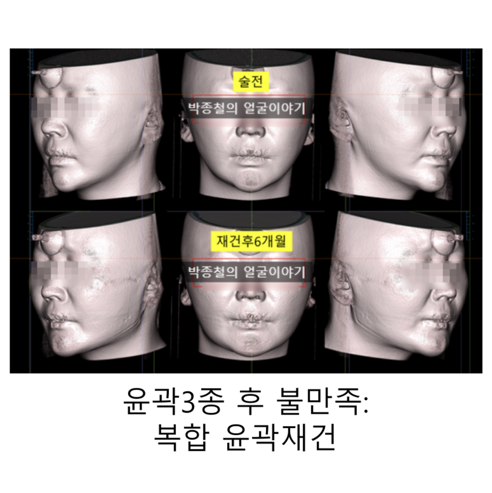
3-Year Post-operative Comparison: CT Analysis

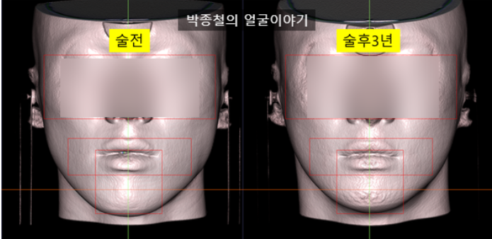
A noticeable reduction in facial length and width is observed (red rectangle in the image). Interestingly, the neck appears thicker. The 45-degree lateral view clearly shows a decrease in facial length (red line).


In the lateral image, it can be seen that the mandibular angle (jaw angle) has moved slightly upward compared to before the surgery, and the chin has become shorter vertically and protrudes forward.


3 Years After Zygoma Reduction: How Has the Bone Changed?
Frontal CT: No significant changes in bone structure are observed. Zygomatic width remains stable.

45-degree CT: Smooth union of the osteotomy site (red arrow).

CT Overlap: Minimal widening of the zygomatic bone is observed, barely noticeable to the naked eye.


Lateral Zygoma: Bone coverage over the metal plate is evident (red arrow).

Lateral Zygoma Reduction and Step-off
This case demonstrates that a step-off (irregularity) after lateral zygoma reduction (red dotted line) can smooth out over time without additional bone shaving

The 3-year follow-up shows a smooth contour in the corresponding soft tissue area.

Long-term Safety and Stability
The 3-year CT analysis confirms the stability of the zygomatic bone and proper healing of the surgical sites, demonstrating the long-term safety of male zygomatic reduction.
Conclusion
This case study provides valuable insights into the long-term skeletal changes following male zygomatic reduction. It highlights the natural smoothing of step-offs in lateral zygoma reduction over time, potentially reducing the need for additional procedures.


Comments