Male Facial Contouring Proven by 3D CT: An 8-Month Before & After of Correcting Facial Asymmetry as an Alternative to Two-Jaw Surgery
- Dr. Park

- Sep 8, 2025
- 4 min read
Recently, a growing number of male patients have visited our clinic seeking to correct facial asymmetry. While many understand that orthognathic (two-jaw) surgery is the most fundamental solution, they often look for alternatives due to the long recovery period and the intensity of the procedure. In such cases, male facial contouring surgery—specifically a 3-point procedure that addresses the jaw, cheekbones, and chin—can be a highly satisfactory alternative.
This article presents an objective, data-driven case study of a male patient who underwent a 3-point facial contouring procedure. Through a precise 3D CT analysis, we identified the primary cause of his facial asymmetry and will now share the detailed 8-month facial contouring before and after results.
Pre-Operative 3D CT Analysis: Identifying the Cause of Facial Asymmetry
Upon his initial visit, the patient's facial asymmetry was clearly visible to the naked eye. To determine the exact cause, we performed a 3D CT scan. The analysis revealed that his right mandibular angle (square jaw bone) protruded outwards more than the left, and his chin tip was also deviated from the centerline to the right.
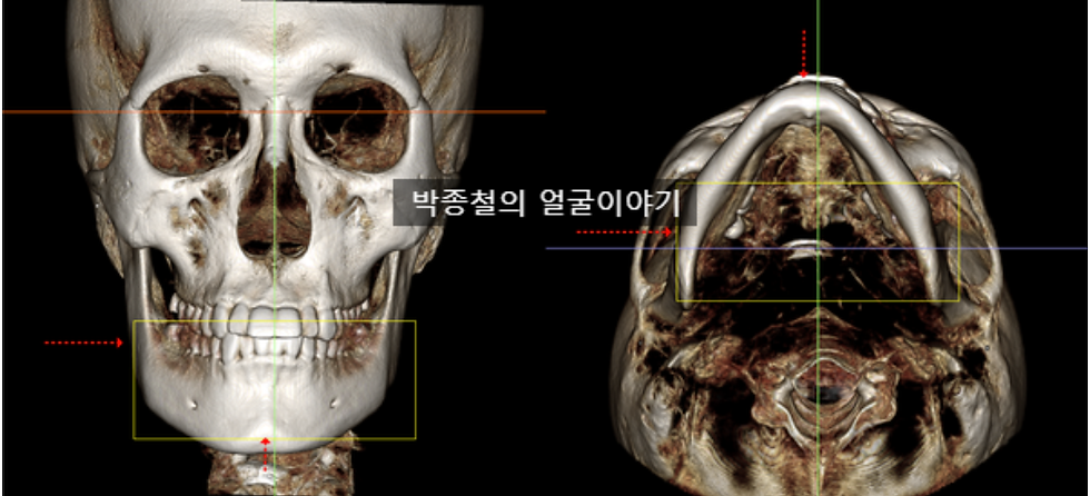
While orthognathic surgery would be the ideal method for correcting such skeletal asymmetry, the patient opted for male facial contouring surgery due to time constraints related to his career and personal concerns about the procedure. He made this decision with a full understanding that minor asymmetry might remain post-surgery.
A Customized Male Facial Contouring Plan Based on Precise CT Analysis
Based on the detailed 3D CT scan results, we established a surgical plan tailored to the patient's unique bone structure.
Mandible (Jaw) Surgery: The plan was to maintain the overall angle of the jaw while performing a greater cortical bone resection on the right side—the main source of the asymmetry—to achieve maximum possible symmetry.
Zygoma (Cheekbone) Surgery: The amount of bone reduction was applied differently to the left and right sides according to their projection. The resected bones were then securely fixed with metal plates to ensure stability.
Genioplasty (Chin) Surgery: A differential resection was planned to adjust the chin, moving its tip back to the facial midline to correct the deviation.
8 Months After Facial Contouring: Objective Skeletal Changes Confirmed by 3D CT
Eight months post-surgery, a follow-up 3D CT scan was taken to objectively compare the skeletal changes from before and after the facial contouring surgery.
Frontal CT Changes

The most noticeable change in the frontal view was the reduction in the width of the right jaw. As planned, the reduction on the right side created a better balance with the left, confirming a significant improvement in symmetry.
Lateral and Axial CT Changes

The lateral and axial CT scans further confirmed that only the planned areas were precisely resected. The reduction in the width of the right jaw was especially clear from the axial view.
How Skeletal Changes Affect Overall Appearance: A Soft Tissue Analysis
To verify how the skeletal changes translated to the patient's actual appearance, we superimposed the before and after photos taken under identical conditions to analyze the changes in the soft tissue (skin, muscle).

The patient had a very low body fat percentage (under 15%) before the surgery, and his weight remained stable afterward. Generally, individuals with lower body fat tend to experience faster dissipation of swelling. This patient also experienced a quick recovery and was able to appreciate the results of his surgery relatively early.

The changes in soft tissue were most apparent from the side, particularly on the right where more cortical bone was resected. This demonstrates that cortical bone resection not only reduces facial width from the front but also plays a crucial role in refining the contour from the side profile.
Conclusion: Successful Male Facial Contouring Begins with a Precise Diagnosis
Male facial contouring surgery is not simply about shaving down bone. The core of a successful procedure lies in scientifically analyzing an individual's unique facial bone structure and the root cause of asymmetry using advanced tools like 3D CT scans. Based on this analysis, the goal is to achieve the most harmonious and balanced result.
This case study demonstrates how a meticulously planned 3-point contouring surgery can be an effective alternative for male patients with facial asymmetry who are hesitant about undergoing orthognathic surgery. The objective facial contouring before and after data speaks for itself.
If you are considering facial contouring surgery, I recommend that you do not make a decision based on appearance alone. The first step toward a satisfactory result is to seek a thorough consultation with an experienced oral and maxillofacial surgeon and receive a precise diagnosis based on a 3D CT analysis to determine the most suitable surgical plan for you.


Comments