Facial Contouring for Asymmetry: A 2.5-Year 3D CT Analysis of a Square Jaw Surgery Case & The Truth About Skin Sagging
- Dr. Park

- Jul 10, 2025
- 4 min read
Updated: Jul 12, 2025
Hello, I am Dr. Park Jong-chul, an Oral and Maxillofacial Surgeon.
Perfect facial symmetry is rare. When asymmetry exceeds a certain degree, many people begin to consider options for improvement. While severe skeletal asymmetry may require double-jaw surgery, it is not the necessary solution for all cases.
For patients like the one in this case study, who had a 3mm difference between the left and right sides of their jaw, facial contouring surgery can achieve highly satisfactory results. This case is particularly informative because it involves an objective analysis of both skeletal and soft tissue changes over a long-term period of 2.5 years post-surgery, using high-resolution 3D CT scans. This in-depth analysis is made possible by my specialized CT equipment, and I am sharing this data to provide clear, evidence-based information and alleviate common patient concerns.
Pre-operative Analysis: Identifying the Cause of Asymmetry
A precise analysis of the patient's pre-operative condition revealed that while the chin's centerline was well-positioned, the right mandibular angle (square jaw) was longer and wider than the left. This discrepancy was the primary cause of the facial asymmetry, which was clearly observable not only from the front but also more distinctly from the side and in mirrored images.


A Comprehensive Surgical Plan for Asymmetry Correction
To improve the patient's asymmetry and create a more harmonious and refined facial structure, the following detailed surgical plan was established:
Square Jaw Reduction (Mandibular Angle Resection): A "differential resection" was performed, meaning different amounts of bone were removed from each side. A larger portion of the more developed right jaw angle was resected, along with an additional reduction of the outer cortical bone, to achieve symmetry from a frontal view.
Genioplasty (T-Osteotomy): To create a slimmer chin line, a 5mm-wide segment of bone was removed from the center of the chin tip. The two sides were then joined together. The plan was specifically designed to resect more of the right mandibular body during this process, further correcting the overall jawline imbalance.
Zygoma Reduction (Cheekbone Reduction): To reduce the overall width of the face and create a softer profile, both cheekbones were moved medially. (Bone resection: 3mm on both sides; 45-degree posterior movement: 2mm; lateral zygoma reduction: 7mm).
2.5 Years Post-Surgery: An Objective, Data-Driven Review
The patient maintained a stable physical condition for 2.5 years following the surgery, with a negligible weight change (1 kg loss) and a minor increase in body fat percentage (0.9%). This is a crucial point, as it allows us to analyze the soft tissue changes as a direct result of the surgery itself.
Skeletal Changes: Symmetrical Reduction Achieved as Planned
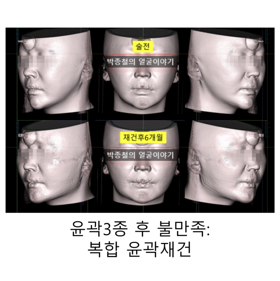
A comparison of the pre- and post-operative 3D CT scans clearly shows that the mandibular angle and zygoma were symmetrically and smoothly reduced according to the surgical plan, without excessive resection. This is objective evidence that the surgery was performed with high precision.

Soft Tissue Changes: The Truth About "Skin Sagging" After Facial Contouring
Let's address the most common patient concern—skin sagging—by examining the actual soft tissue changes 2.5 years after surgery.

To state the conclusion first: vertical sagging, where the skin droops downwards due to gravity, does not occur after bone resection. The soft tissues, including skin and muscle, adhere firmly to the new surface of the resected bone over time.
To illustrate this, we have overlaid the pre-operative soft tissue contour (yellow line) onto the post-operative CT cross-section. As you can see, there is no area where the tissue has sagged below the original contour line.

So, why do some people feel a sense of "sagging"?
This perception is not due to the tissue "sagging" downwards, but rather a subtle "redistribution" of the remaining soft tissue volume, which settles onto the new, smaller bone structure. In the analysis image showing soft tissue thickness, the areas marked in green indicate where the tissue has become thicker compared to before the surgery.

However, this volume shift is typically very slight and not perceived as "sagging" by most patients. In this case, there was no noticeable sagging observed in the patient's soft tissue imaging.

In-depth Soft Tissue Analysis: Changes in the Masseter Muscle and Buccal Fat
Post-surgical satisfaction is determined by the harmony between bone and soft tissue. We will now look closely at the changes in the masseter (chewing) muscle and the buccal fat pad, which significantly influence mid-face volume.
Masseter Muscle: Typically, after square jaw surgery, the masseter muscle is redistributed anteriorly even as its overall volume decreases. However, this patient's case was unique in that there was minimal forward redistribution; instead, there was a more direct reduction in the muscle's volume. This means the volumizing effect on the mid-face from muscle redistribution was less pronounced.

사각턱 수술전 연조직의 경계는 노란선으로, 심부볼, 교근의 경계는 빨간선 
원본이미지 Buccal Fat: This patient did not undergo buccal fat removal. As expected, the CT analysis confirmed that the soft tissue adjacent to the buccal fat pad became slightly thicker post-surgery. This highlights the importance of accurately diagnosing the buccal fat pad during surgical planning; for some patients, combining the procedure with buccal fat removal can be crucial for achieving a more refined and satisfactory result.

안면윤곽수술후 심부볼의 영향

Conclusion: A Comprehensive Approach is Key to Successful Facial Contouring
This long-term case study provides us with a clear conclusion.
The "skin sagging" commonly feared after facial contouring surgery is not a vertical drop but a natural process of soft tissue redistribution onto a new skeletal framework. Therefore, a successful surgical outcome depends not just on planning the amount of bone resection, but on a comprehensive approach that anticipates and accounts for these dynamic soft tissue changes.
Ultimately, the best results are achieved when the surgeon's accurate diagnosis and rich experience are combined with the patient's dedicated post-operative efforts. Particularly for patients with a higher-than-average body fat percentage before surgery, active efforts to reduce body fat afterward can dramatically enhance the final outcome.
I hope this objective, data-driven review helps you make an informed and wise decision.

Facial Contouring for Asymmetry


Comments