Revision Surgery for Mandibular Angle Asymmetry: A Bone Reconstruction Approach
- Dr. Park

- May 6, 2025
- 3 min read
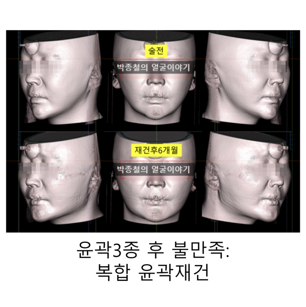
Mandibular angle reduction surgery is a common procedure to improve facial contours. However, some patients may experience dissatisfaction due to over-resection or asymmetry. In such cases, revision surgery should be considered not as a means of further reduction, but as a restoration of lost volume and symmetry.
Understanding Skeletal Asymmetry Before Revision Surgery
Before planning revision mandibular angle surgery, it is essential to understand the concept of skeletal asymmetry. Perfect facial symmetry is rare. Variations in cranial bone width and shape are common, and any attempt to restore balance must begin with objective analysis using 3D computed tomography (CT) data.


To achieve this, a standardized coordinate system must be established. This is similar to aligning a subject’s pose when taking an ID photo—facial skeletal data must be oriented to the frontal, axial, and sagittal planes based on anatomical landmarks. These references may include the interpupillary line, the nasion-midline, or the zygomaticofrontal suture.

Using this coordinate system, the degree and pattern of mandibular asymmetry can be quantitatively analyzed. For instance, the frontal ramus inclination or external oblique ridge (EOR) angle may guide the definition of a balanced appearance. A clear surgical plan is derived from this objective evaluation.
Approaching Revision Surgery from a Reconstructive Perspective
When people think of revision mandibular angle surgery, they often assume it involves additional bone reduction. However, in cases where the mandibular angle was excessively resected during a previous procedure, restoring the lost volume becomes the primary surgical goal.
Therefore, revision surgery from a reconstructive perspective aims to restore a balanced jaw contour and improve facial projection. The surgical approach should focus not on further reduction but on volumetric enhancement and symmetry correction. Selecting the appropriate reconstructive material is essential to achieving these goals.
Autogenous Bone Graft vs. Alloplastic Implant
Two main methods are used for this purpose:
Autogenous Bone Grafting
Bone is harvested from the patient’s own body, usually the iliac crest, and grafted to the mandibular angle area. While biologically compatible, this technique carries risks of donor-site complications such as scarring, pain, altered sensation, and infection. Furthermore, the long-term volume retention of grafted bone is difficult to predict due to resorption.

Kook, I., You, J., Kim, D.H. et al. A retrospective cohort study of autogenous iliac strut bone grafting in large bone defects of the lower extremity. Sci Rep 14, 6059 (2024). https://doi.org/10.1038/s41598-024-56726-7 Alloplastic Implant Augmentation
A custom-made implant—often based on the patient's 3D CT data—is used to restore the contour. This method allows for precise volume and shape recovery. It avoids the need for additional surgery at a donor site, and the final result is often more predictable.
Why Titanium Implants Are a Reliable Choice
Among various implant materials, titanium stands out for its biocompatibility and durability. Titanium has long been used in orthopedic and dental implants due to the following advantages:
Excellent Biocompatibility: It rarely causes adverse reactions or inflammation.
High Strength and Stability: It maintains shape over time with minimal risk of deformation.
Precision Manufacturing: Using 3D printing and CAM technologies, titanium implants can be tailored to each patient’s anatomy with high accuracy.

Ensuring Precise Placement Through 3D Simulation and Surgical Guides
Even a perfectly designed implant may not yield optimal results if not placed precisely. Small positional deviations can significantly affect facial symmetry and aesthetics. Therefore, I utilize the following protocol:
3D Surgical Simulation: Preoperative simulation based on CT data helps define optimal placement, screw fixation, and nerve safety.
Custom Surgical Guides: Patient-specific guides are fabricated to assist in placing the implant in the planned position, minimizing intraoperative error and maximizing surgical accuracy.

Conclusion
Revision mandibular angle surgery demands careful planning and precise execution. Especially when the goal is to restore symmetry and volume after over-resection, objective assessment using 3D imaging and the use of biocompatible, custom titanium implants can lead to predictable and satisfactory outcomes. My approach integrates digital planning and surgical guides to deliver consistent results and improve patient satisfaction.
If you have concerns about previous mandibular angle surgery results, I encourage you to consult with a qualified specialist for an individualized evaluation.


Comments